
Diagnostic imaging
3D Meta-Vu Skin Analysis
The consultation starts with a photograph you can argue with.
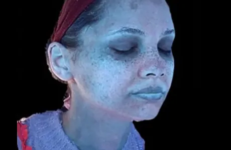
Meta-Vu photographs the face in 3D with light positioned around its contours, then separates the image into texture, pigment, pores, wrinkles, tone, dark circles, redness, melanin, sebum, acne bacteria and predicted lines. It replaces opinion with something you can both look at.
- Works on
- Full-face diagnostic
- Sensation
- None
- Downtime
- None
- Typically lasts
- Repeated to track change
Diagnostic imaging
What it addresses
Read the pigment first. Then choose the wavelength.
- 01Pigment, redness and pore condition mapped objectively
- 02An analysis you can compare against, not just describe
- 03A treatment plan built from your own skin data
- 04Before-and-after imaging on the same lighting rig
How it works
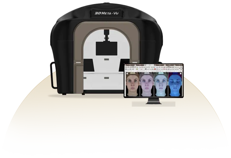
3D capture
Light is placed around the curve of the face so the sides read as accurately as the front.
Eleven layers in one sitting
Everything from sebum to predicted wrinkle lines is captured at once, so nothing has to be estimated.
Easy to read
Image-based analysis means you can see the reasoning behind a recommendation rather than take it on trust.
Every plan is decided in consultation. Suitability, sequence and dosage differ from person to person.
The instrument
3D Meta-Vu Skin Analysis
Diagnostic imaging
Also in this group